FAQ: How do I know that my HMW DNA has attached to glass beads in the Monarch HMW DNA Extraction Kits?

When samples contain >10 µg of HMW DNA, which is common when working with ‘Standard Input’ amounts, the binding of the DNA to the beads is visible and will be seen as a cloudy, opaque layer covering the beads. The beads will always stick together after this amount of DNA is bound to them. When inversions are done manually using ‘Standard Input’ amounts, the binding process can be seen and followed. After 5 inversions, the cloud-like DNA becomes visible, and in the next 10 inversions, it connects to the beads and starts wrapping around them. At this point, the solution remains viscous. Once the DNA is completely wrapped around the beads during the remaining 10-15 inversions, the solution will no longer be viscous. ‘Low Input’ samples typically yield between 1-10 µg of HMW DNA and the binding of the DNA may be more difficult to follow. If only 1-5 µg of DNA is present, it will likely not be possible to see the DNA binding to the beads; the beads will seem to move freely and independently in solution. When there is between 5-10 µg of DNA, it is difficult to see the DNA binding to the beads, but the bound DNA will often cause the beads to stick together.
  1. Monarch-HMW-Video_Cell_Blood_1920

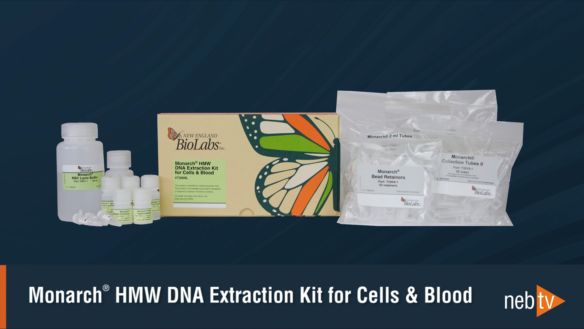
    Monarch® HMW DNA Extraction from Cell & Blood: Protocol Overview

    In this video, we walk through the protocol for extraction of high molecular weight DNA (HMW DNA) from cultured cells.

  2. Monarch-HMW-Video_Tissue_1920

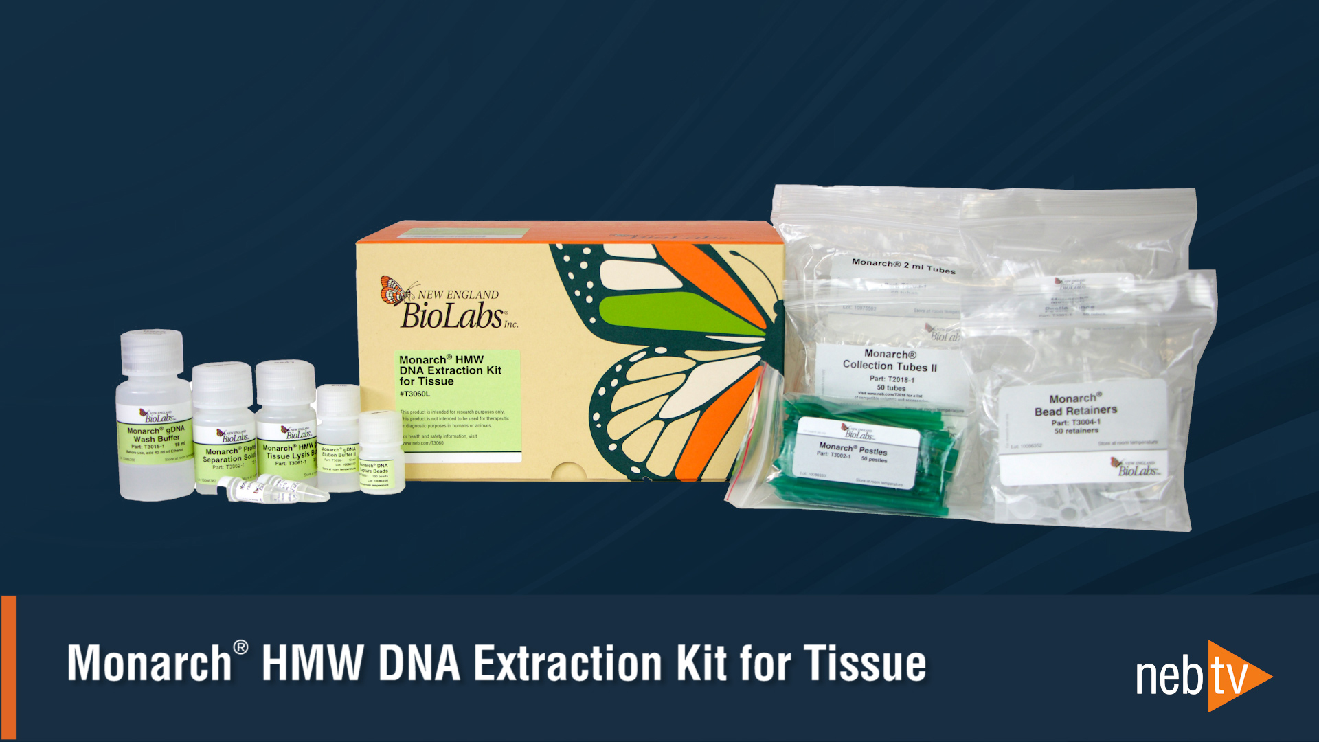
    Monarch® HMW DNA Extraction from Tissue: Protocol Overview

    In this video, we walk through the protocol for extraction of high molecular weight DNA (HMW DNA) from tissues.