Home
Applications
Glycobiology & Proteomics
Biotherapeutics and Antibody Analysis
N-glycan Composition Profiling
N-glycan Composition Profiling
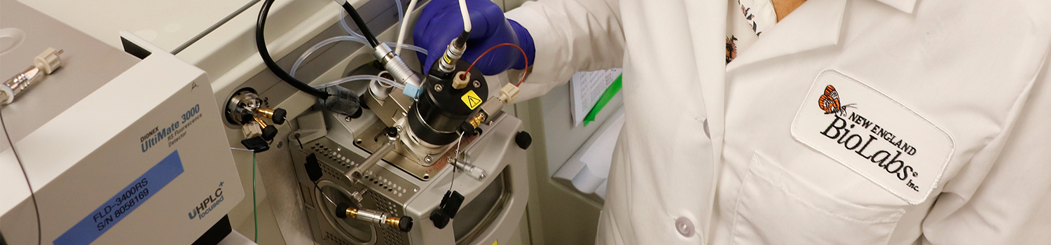
Return to Biotherapeutics and Antibody AnalysisDuring the production of recombinant glycoproteins, even slight process variations might lead to changes in glycosylation. Unlike the primary sequence of a protein (that can be engineered at the DNA level), the structure of glycans is not encoded in the genome. The cells’ metabolic rate, the availability of nutrients, and the fermentation conditions effect the levels of precursors (nucleotide sugars) and biosynthetic enzymes, which ultimately determine the exact glycan composition. Therefore, it is critical to monitor glycosylation during the manufacturing of a biological drug.
- Modern quality-by-design (QbD) and continuous manufacturing principles demand glycan analysis methods that are fast, high-throughput, accurate, reproducible, and flexible.
- To overcome time constraints in N-glycan release by PNGase F, NEB developed a novel enzyme, Rapid™ PNGase F (P0710), for deglycosylation of antibodies and fusion proteins in minutes. Rapid™ PNGase F completely removes all N-glycan sites without bias, allowing the measurement of all species (regardless of abundance) with accuracy.
- N-glycans have to be labeled with fluorescent tags for sensitive detection and quantitation during liquid chromatography coupled with fluorescence detection (LC-FLD). These fluorescent tags might also assist ionization for mass spectrometry (MS), or bear charges for capillary electrophoresis (CE). Commonly used tags are 2-aminobenzamide (2AB), 2-aminobenzoic acid (2AA), procainamide (PCA), and 8-Aminopyrene-1,3,6-trisulfonic acid (APTS, which is used in CE).
- These tags are introduced via reductive amination, in the presence of an acid catalyst and a reducing agent.
- Detailed structural analysis of released N-glycans is possible by a combination of LC-MS analysis, which identifies N-glycans by retention time and exact mass, coupled with an exoglycosidase array analysis (N-Glycan Sequencing Kit E0577s).